Abstract
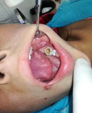
A clinical case of a 9-year-old patient who seeks care for elevated lesions of the oral mucosa is presented. A disease caused by HPV was diagnosed. This case might potentially involve child abuse due to modes of disease transmission. On clinical examination, an ulcerated papillary lesion of approximately 0.5 cm was observed in the right lip corner. Multiple raised warty lesions were seen on both cheeks and the lower labial mucosa. We conclude about the importance of a good clinical history and a complete oral examination to implement the right treatment and follow-up of pathologies.
References
1. Dunne EF, Nielson CM, Stone KM, Markowitz LE, Giuliano AR. Prevalence of HPV infection among men: A systematic review of the literature. J. Infect. Dis. 2006;194 (8): 1044-57.
2. Dunne EF, Unger ER, Sternberg M, McQuillan G, Swan DC, Patel SS, Markowitz LE. Prevalence of HPV infection among females in the United States. J Am Med Assoc 2007; 297(8): 813–9.
3. López A, Basurto C, Salazar R. VPH en cavidad oral: condiloma. Rev Tamé 2019; 7(21): 838-41.
4. Estrada G, Márquez M, González E, Nápoles M, Ramón R. Infección por virus del papiloma humano en la cavidad bucal. Medisan 2015; 19(3): 55-78.
5. Betz SJ. HPV-related papillary lesions of the oral mucosa: A review. Head Neck Pathol. 2019;13(1): 80-90. doi: 10.1007/s12105-019-01003-7.
6. Cháirez Atienzo P, Vega Memíje ME, Zambrano Galván G, García Calderón AG, Maya García IA, Cuevas González JC. Presencia del virus papiloma Humano en la cavidad oral: Revisión y actualización de la Literatura. Int. J. Odontostomatol. 2015; 9(2) :233–8.
7. González M, Suarez R, Canul J, Conde L, Eljure N. Multifocal epithelial hyperplasia in a community in the Mayan area of Mexico. Int. J. Dermatol. 2011; 50(3): 304-9. doi:10.1111/j.1365-4632.2010.04718.
8. Yarmuch P, Chaparro X, Fischer C, Benveniste S. Enfermedad de Heck: A propósito de un caso. Rev. Chilena Dermatol.2012; 28(4):431–4.
9. Obalek S, Jablonska S, Favre M, Walczak L, Orth G. Condylomata acuminata in children: frequent association with human papillomaviruses responsible for cutaneous warts. J. Am. Acad. Dermatol. 1990; 23 (2 Pt 1):.205-13.
10. Bennett LK, Hinshaw M. Heck’s disease: diagnosis and susceptibility. Pediatr. Dermatol. 2009; 26(1): 87-9.
11. González LV, Gaviria AM, Sanclemente G, Rady P, Tyring SK, Carlos R, Correa LA, Sanchez GI. Clinical, histopathological and virological findings in patients with focal epithelial hyperplasia from Colombia. Int. J. Dermatol. 2005; 44(4): 274-9.
12. Harris AM, Van Wyk CW. Heck’s disease (focal epithelial hyperplasia): a longitudinal study. Community Dent. Oral Epidemiol. 1993; 21(2): 82-5.
13. Dos Santos PJ, Bessa CF, De Aguiar MC, Do Carmo MA. Cross-sectional study of oral mucosal conditions among a central Amazonian Indian community, Brazil. J. Oral Pathol. Med. 2004;3(1):.7-12.
14. Ledesma-Montes C, Vega-Memije E, Garcés-Ortiz M, Cardiel-Nieves M, Juárez-Luna C. Hiperplasia multifocal del epitelio: Reporte de nueve casos. Med. Oral Patol. Oral Cir. Bucal. 2005 ;10(5):.394–401.
15. Nartey NO, Newman MA, Nyako EA. Focal epithelial hyperplasia: report of six cases from Ghana, West Africa. J. Clin. Pediatr. Dent. 2002; 27(1): 63-6.
16. Luciano R, Oviedo J. Virus del papiloma humano y cáncer bucal. Acta Odontol. Venez., 2013; 51(1): 1-3.


