Abstract
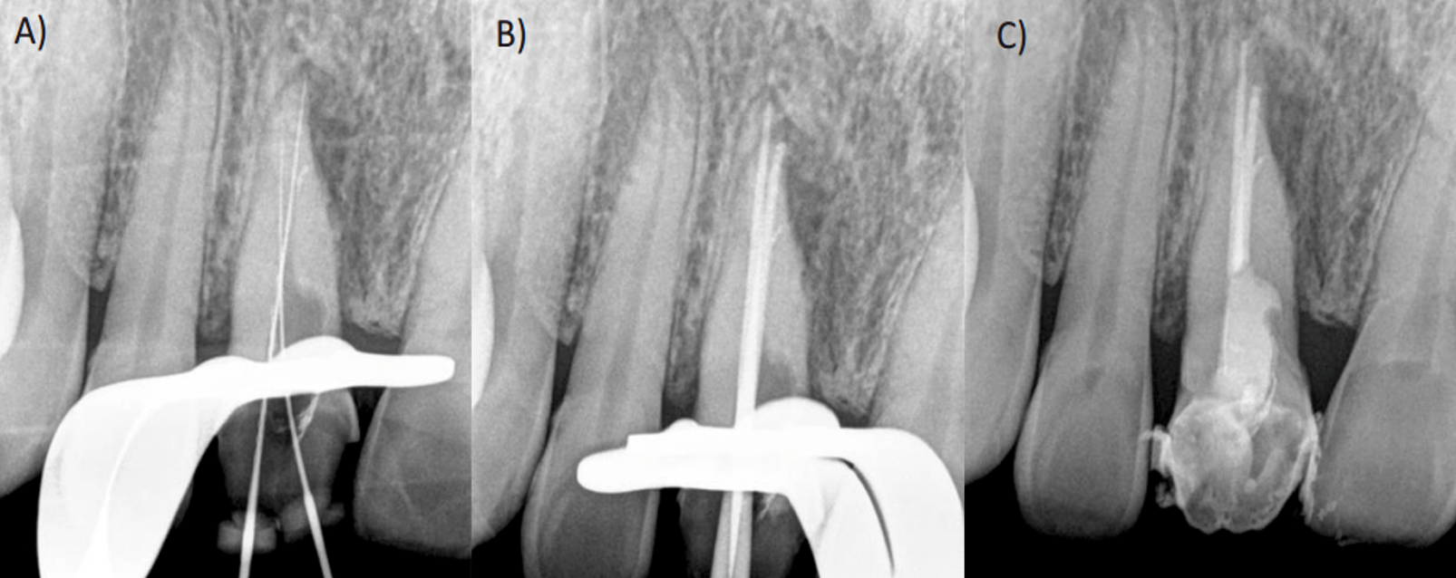
The failure in root canal treatment can lead to the loss of the dental organ. Various factors have been reported as causes
of failure in endodontic therapy. Among these factors, omitted canals have a greater influence, as they allow colonization and
multiplication of bacteria within the root canal. Abnormal anatomical variations can increase the chances of failure due to the
inability to diagnose accurately. In these cases, cone-beam computed tomography has proven to be of great assistance in their
interpretation
References
Graves DT, Oates T, Garlet GP. Review of osteoimmunology and the host response in endodontic and periodontal lesions. J Oral Microbiol. 2011;3.
Berman LH, Hargreaves KM. Cohen’s pathways of the pulp-e-book: Elsevier Health Sciences; 2020.
Rotstein I, Ingle JI. Ingle’s endodontics: PMPH USA; 2019.
Teixeira QE, Ferreira DC, da Silva AMP, Goncalves LS, Pires FR, Carrouel F, et al. Aging as a Risk Factor on the Immunoexpression of Pro-Inflammatory IL-1beta, IL-6 and TNF-alpha Cytokines in Chronic Apical Periodontitis Lesions. Biology (Basel). 2021;11(1).
Thuller K, Armada L, Valente MI, Pires FR, Vilaca CMM, Gomes CC. Immunoexpression of Interleukin 17, 6, and 1 Beta in Primary Chronic Apical Periodontitis in Smokers and Nonsmokers. J Endod. 2021;47(5):755-61.
Popovska L, Dimova C, Evrosimoska B, Stojanovska V, Muratovska I, Cetenovic B, et al. Relationship between IL-1ȕproduction and endodontic status of human periapical lesions. Vojnosanitetski pregled. 2017;74(12):1134-9.
Boersma B, Jiskoot W, Lowe P, Bourquin C. The interleukin-1 cytokine family members: Role in cancer pathogenesis and potential therapeutic applications in cancer immunotherapy. Cytokine Growth. Factor Rev. 2021;62:1-14.
Siqueira JF, Jr., Rocas IN. Present status and future directions: Microbiology of endodontic infections. Int Endod J. 2022;55 Suppl 3:512-30.
Nair PN. On the causes of persistent apical periodontitis: a review. Int Endod J. 2006;39(4):249-81.
Takahashi K. Microbiological, pathological, inflammatory, immunological and molecular biological aspects of periradicular disease. Int Endod J. 1998;31(5):311-25.
Orstavik D. Essential endodontology: prevention and treatment of apical periodontitis: John Wiley & Sons; 2020.
Boutsioukis C, Arias-Moliz MT. Present status and future directions - irrigants and irrigation methods. Int Endod J. 2022;55 Suppl 3(Suppl 3):588-612.
Chaniotis A, Ordinola-Zapata R. Present status and future directions: Management of curved and calcified root canals. Int Endod J. 2022;55 Suppl 3:656-84.
Vertucci FJ. Root canal morphology and its relationship to endodontic procedures. Endodontic Topics. 2005;10(1):3-29.
Karabucak B, Bunes A, Chehoud C, Kohli MR, Setzer F. Prevalence of Apical Periodontitis in Endodontically Treated Premolars and Molars with Untreated Canal: A Cone-beam Computed Tomography Study. J Endod. 2016;42(4):538-41.
Mustafa M, Almuhaiza M, Alamri HM, Abdulwahed A, Alghomlas ZI, Alothman TA, et al. Evaluation of the causes of failure of root canal treatment among patients in the City of Al-Kharj, Saudi Arabia. Niger J Clin Pract. 2021;24(4):621-8.
Baruwa AO, Martins JNR, Meirinhos J, Pereira B, Gouveia J, Quaresma SA, et al. The Influence of Missed Canals on the Prevalence of Periapical Lesions in Endodontically Treated Teeth: A Crosssectional Study. J Endod. 2020;46(1):34-9 e1.
Pessotti VP, Jiménez-Rojas LF, Alves FRF, Rôças IN, Siqueira JF, Jr. Post-treatment apical periodontitis associated with a missed root canal in a maxillary lateral incisor with two roots: A case report. Aust Endod J. 2022.
Siqueira Junior JF, Rocas IDN, Marceliano-Alves MF, Perez AR, Ricucci D. Unprepared root canal surface areas: causes, clinical implications, and therapeutic strategies. Braz Oral Res. 2018;32(suppl 1):e65.
Costa F, Pacheco-Yanes J, Siqueira JF, Jr., Oliveira ACS, Gazzaneo I, Amorim CA, et al. Association between missed canals and apical periodontitis. Int Endod J. 2019;52(4):400-6.
Patel S, Dawood A, Whaites E, Pitt Ford T. New dimensions in endodontic imaging: part 1. Conventional and alternative radiographic systems. Int Endod J. 2009;42(6):447-62.
Ball RL, Barbizam JV, Cohenca N. Intraoperative endodontic applications of cone-beam computed tomography. J Endod. 2013;39(4):548-57.
Cotti E, Schirru E. Present status and future directions: Imaging techniques for the detection of periapical lesions. Int Endod J. 2022;55 Suppl 4:1085-99.
Setzer FC, Lee SM. Radiology in Endodontics. Dent Clin North Am. 2021;65(3):475-86.
Decurcio DA, Bueno MR, Silva JA, Loureiro MAZ, Damião Sousa-Neto M, Estrela C. Digital Planning on Guided Endodontics Technology. Braz Dent J. 2021;32(5):23-33.
Setzer FC, Kratchman SI. Present status and future directions: Surgical endodontics. Int Endod J. 2022;55 Suppl 4:1020-58.
Ozcan G, Sekerci AE, Cantekin K, Aydinbelge M, Dogan S. Evaluation of root canal morphology of human primary molars by using CBCT and comprehensive review of the literature. Acta Odontol Scand. 2016;74(4):250-8.
Yan Y, Li J, Zhu H, Liu J, Ren J, Zou L. CBCT evaluation of root canal morphology and anatomical relationship of root of maxillary second premolar to maxillary sinus in a western Chinese population. BMC Oral Health. 2021;21(1):358.
Glickman GN. AAE Consensus Conference on Diagnostic Terminology: background and perspectives. J Endod. 2009;35(12):1619-20.
Ahmed HMA, Dummer PMH. A new system for classifying tooth, root and canal anomalies. Int Endod J. 2018;51(4):389-404.
Dexton AJ, Arundas D, Rameshkumar M, Shoba K. Retreatodontics in maxillary lateral incisor with supernumerary root. J Conserv Dent. 2011;14(3):322-4.
Levin A, Shemesh A, Katzenell V, Gottlieb A, Ben Itzhak J, Solomonov M. Use of Cone-beam Computed Tomography during Retreatment of a 2-rooted Maxillary Central Incisor: Case Report of a Complex Diagnosis and Treatment. J Endod. 2015;41(12):2064-7.
Kang M, Kim E. Unusual morphology of permanent tooth related to traumatic injury: a case report. J Endod. 2014;40(10):1698-701.
Coutinho T, Lenzi M, Simoes M, Campos V. Duplication of a permanent maxillary incisor root caused
by trauma to the predecessor primary tooth: clinical case report. Int Endod J. 2011;44(7):688-95.
Kaufman AY, Keila S, Wasersprung D, Dayan D. Developmental anomaly of permanent teeth related to traumatic injury. Endod Dent Traumatol. 1990;6(4):183-8.
Genovese FR, Marsico EM. Maxillary Central Incisor with Two Roots: A Case Report. Journal of Endodontics. 2003;29(3):220-1.
Jafari Z, Kazemi A, Shiri Ashtiani A. Endodontic Management of a Two-Rooted Maxillary Central Incisor Using Cone-Beam Computed Tomography: A Case Report. Iran Endod J. 2022;17(4):220-2.
Mahadevan M, Paulaian B, Ravisankar SM, Arvind Kumar A, Nagaraj NJ. Endodontic Management of Maxillary Central Incisor with Two Roots, and Lateral Incisor with a C-shaped Canal; A Case Report. Iran Endod J. 2023;18(2):104-9.
Orhan EO, Dereci O, Irmak O. Endodontic Outcomes in Mandibular Second Premolars with Complex Apical Branching. J Endod. 2017;43(1):46-51.
Patel S, Brown J, Semper M, Abella F, Mannocci F. European Society of Endodontology position statement: Use of cone beam computed tomography in Endodontics: European Society of Endodontology (ESE) developed by. Int Endod J. 2019;52(12):1675-8.

This work is licensed under a Creative Commons Attribution-NonCommercial 4.0 International License.

