

Palabras clave
Endodoncia
Diagnóstico
Bioética
Anomalías dentales
Comportamiento de reducción de riesgos
Cómo citar
Resumen
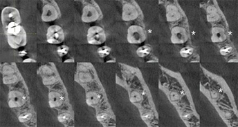
La Tomografía Computarizada de Haz Cónico (CBCT) representa una promisoria herramienta para la clínica odontológica. En Endodoncia, CBCT ofrece tridimensionalidad y resolución imagenológica, potenciando el diagnóstico de diferentes condiciones patológicas. Sin embargo, su limitación por sobreexposición a radiación, ha llevado a directrices que recomiendan cautela para su indicación. Se presenta un caso infrecuente de un molar mandibular con una sola raíz y canal, y las circunstancias de uso de CBCT. Mujer de 48 años es derivada para endodoncia del primer molar mandibular izquierdo permanente. El examen radiográfico preoperatorio demostró el hallazgo inusual de un canal centrado en una única raíz. Con la información apropiada y el consentimiento de la paciente, la indicación de CBCT favoreció su diagnóstico y tratamiento. CBCT no debería ser utilizada rutinariamente para estos fines, pero podría justificarse en casos "fronterizos". Se discuten su indicación e implementación clínica, siguiendo actuales recomendaciones y directrices.
Referencias
2. Patel S, Durack C, Abella F, Shemesh H, Roig M, Lemberg K. Cone beam computed tomography in Endodontics - a review. Int. Endod, J. 2015; 48(1): 3-15. doi: 10.1111/iej.12270.
3. Gulsahi A, Ates U, Tirali RE, Cehreli SB. Use of cone-beam computed tomography in diagnosis of an otherwise undetected periapical lesion in an anomalous tooth. Oral Radiol. 2014; 30: 111-4. doi: 10.1007/s11282-013-0130-8.
4. AAE and AAOMR Joint Position Statement: Use of Cone Beam Computed Tomography in Endodontics 2015 Update. J. Endod. 2015; 41(9): 1393-6. doi: 10.1016/j.joen.2015.07.013.
5. Noffke CE, Farman AG, Nel S, Nzima N. Guidelines for the safe use of dental and maxillofacial CBCT: a review with recommendations for South Africa. SADJ. 2011; 66(6): 262, 4-6.
6. Kiljunen T, Kaasalainen T, Suomalainen A, Kortesniemi M. Dental cone beam CT: A review. Phys. Med. 2015; 31(8): 844-60. doi: 10.1016/j.ejmp.2015.09.004.
7. Sooriaprakas C, Ballal S, Velmurugan N. Mandibular first molar with a single root and single canal. Case Rep. Dent. 2014; 2014: 159846. doi: 10.1155/2014/159846.
8. Filpo-Perez C, Bramante CM, Villas-Boas MH, Húngaro Duarte MA, Versiani MA, Ordinola-Zapata R. Micro-computed tomographic analysis of the root canal morphology of the distal root of mandibular first molar. J. Endod. 2015; 41(2): 231-6. doi: 10.1016/j.joen.2014.09.024.
9. Munavalli A, Kambale S, Ramesh S, Ajgaonkar N. Mandibular first molar with single root and single root canal. J. Conserv. Dent. 2015; 18(4): 346-8. doi: 10.4103/0972-0707.159757.
10. Ioannidis K, Lambrianidis T, Beltes P, Besi E, Malliari M. Endodontic management and cone-beam computed tomography evaluation of seven maxillary and mandibular molars with single roots and single canals in a patient. J. Endod 2011; 37(1): 103-9. doi: 10.1016/j.joen.2010.09.001.
11. Demirbuga S, Sekerci AE, Dinçer AN, Cayabatmaz M, Zorba YO. Use of cone-beam computed tomography to evaluate root and canal morphology of mandibular first and second molars in Turkish individuals. Med Oral Patol Oral Cir Bucal. 2013; 18(4): e737-44. doi: 10.4317/medoral.18473.
12. Raghavendra SS, Napte BD, Desai NN, Hindlekar AN. Single C-shaped canal in mandibular first molar: A case report. J. Conserv Dent. 2015; 18(2): 168-71. doi: 10.4103/0972-0707.153060.
13. Vertucci FJ. Root canal anatomy of the human permanent teeth. Oral Surg Oral Med Oral Pathol Oral Radiol Endodontics. 1984; 58(5): 589-99. doi: 10.1016/0030-4220(84)90085-9.
14. Haridoss S, Swaminathan K, Rajendran V, Rajendran B. Single-rooted primary first mandibular molar. BMJ Case Rep. 2014; 2014. pii: bcr2014206347. doi: 10.1136/bcr-2014-206347.
15. Gaur A, Trivedi HP, Gupta M, Sharma A, Likhyani L, Agarwal M. Mandibular First Molar with Vertucci Type I Canal Configuration diagnosed with the Help of Cone Beam Computed Tomography: A Rare Case Report. J Contemp Dent Pract. 2014; 15(6): 784-7. doi: 10.5005/jp-journals-10024-1618.
16. de Pablo OV, Estevez R, Heilborn C, Cohenca N. Root anatomy and canal configuration of the permanent mandibular first molar: clinical implications and recommendations. Quintessence Int. 2012;43(1):15-27.
17. Torres A, Jacobs R, Lambrechts P, Brizuela C, Cabrera C, Concha G, Pedemonte ME. Characterization of mandibular molar root and canal morphology using cone beam computed tomography and its variability in Belgian and Chilean population samples. Imaging Sci Dent. 2015; 45(2): 95-101. doi: 10.5624/isd.2015.45.2.95.
18. Delgado M, Ramírez LM, Adhikari K, Fuentes-Guajardo M, Zanolli C, Gonzalez-José R, Canizales S, Bortolini MC, Poletti G, Gallo C, Rothhammer F, Bedoya G, Ruiz-Linares A. Variation in dental morphology and inference of continental ancestry in admixed Latin Americans. Am J Phys Anthropol. 2019; 168(3): 438-47. doi: 10.1002/ajpa.23756.
19. Velozo C, Prado VFF, Sousa ISDS, Albuquerque MBA, Montenegro L, Silva S, Silva P, Albuquerque D. Scope of Preparation of Oval and Long-Oval Root Canals: A Review of the Literature. ScientificWorldJournal. 2021; 2021:5330776. doi: 10.1155/2021/5330776.
20. Busquim S, Cunha RS, Freire L, Gavini G, Machado ME, Santos M. A micro-computed tomography evaluation of long-oval canal preparation using reciprocating or rotary systems. Int Endod J. 2015; 48(10): 1001-6. doi: 10.1111/iej.12398.
21. Yendreka VC, Fonseca GM. A "borderline" dental trauma with 12 y of evolution justifying CBCT as diagnostic method. Biomed. Res. 2018; 29(13): 2800-5. doi: 10.4066/biomedicalresearch.29-18-728.
22. Noffke CE, Farman AG, Van der Linde A, Nel S. Responsible use of cone beam computed tomography: minimising medico-legal risks. SADJ. 2013; 68(6): 256, 8-9.
23. Naseri M, Safi Y, Akbarzadeh Baghban A, Khayat A, Eftekhar L. Survey of Anatomy and Root Canal Morphology of Maxillary First Molars Regarding Age and Gender in an Iranian Population Using Cone-Beam Computed Tomography. Iran Endod. J. 2016; 11(4): 298-303. doi: 10.22037/iej.2016.8.
24. Scarfe WC. "All that glitters is not gold": standards for cone-beam computerized tomographic imaging. Oral Surg Oral Med Oral Pathol Oral Radiol Endod. 2011; 111(4): 402-8. doi: 10.1016/j.tripleo.2011.01.006.
25. Rosen E, Taschieri S, Del Fabbro M, Beitlitum I, Tsesis I. The Diagnostic Efficacy of Cone-beam Computed Tomography in Endodontics: A Systematic Review and Analysis by a Hierarchical Model of Efficacy. J. Endod. 2015; 41(7): 1008-14. doi: 10.1016/j.joen.2015.02.021.