

Versões
- 2024-07-12 (2)
- 2021-09-30 (1)
Palavras-chave
Língua
Via Aérea
Maloclusão
Como Citar
Resumo
Esta investigação centrou-se na revisão dos artigos que avaliaram cefalometricamente a posição do osso hióide, a posição linguística e a dimensão da via aérea faríngea de acordo com a maloclusão esquelética, a fim de determinar se existe uma relação entre estes estruturas.
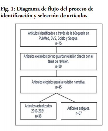
Método: As publicações foram identificadas nas seguintes bases de dados: PubMed, Biblioteca Virtual em Saúde (BVS), Scielo e Scopus. Palavras-chave: Osso hióide, Língua, Via aérea, Maloclusão. Os artigos foram analisados por título, resumo e texto completo, escritos em inglês e espanhol.
Resultados: Foram encontrados 75 artigos, 30 foram eliminados porque não estavam directamente relacionados com o tema. Finalmente, foram seleccionados 45 artigos.
Conclui-se que ainda não existe consenso absoluto sobre a relação entre: a posição do osso hióide, a língua e a dimensão da via aérea superior, de acordo com a maloclusão esquelética.
Referências
Proffit WR. Ortodoncia contemporánea. 4ta Ed. España: Editorial Elsevier; 2008.
Iwasaki T, Suga H, Minami A, Sato H, Hashiguchi M, Tsujii T. Ralationships among tongue volumen, hyoid position, airway volumen, and maxilofacial form in pediatric patients with Class-II, and ClassIII malocclusions. Orthod Craniofac. 2019;22(1):9-15.
McNamara JA. Influence of respiratory pattern on craniofacial growth. Angle Orthod. 1981;51:269-300.
Moss M. The primary role of functional matrices in facial growth. Am J Orthod. 1959;55:566-567.
Kaur S, Rai S, Kaur M. Comparación de la confiabilidad del cefalograma lateral y la tomografía computarizada para la evaluación del espacio de las vías respiratorias. Niger J Clin Pract. 2014;17:629-36.
Nejaim Y, Johan K, Groppo F, Neto F. Evaluation of pharyngeal space and its correlation with mandible and hyoid bone in patients with different skeletal classes and facial types. American Journal of Orthodontics and Dentofacial Orthopedics. 2018;153(6):825-833.
Liégeois F, Albert A, Limme M. Comparison between tongue volume from magnetic resonance images and tongue area from profile cephalograms.Eur J Orthod. 2010; 32(4): 381–6.
Shokri A, Mollabashi V, Zahedi F, Tapak L. Position of the hyoid bone and its correlation with airway dimensions in different classes of skeletal malocclusion using cone-beam computed tomography. Imaging Science in Dentistry. 2020;50:105-15.
Mortazavi S, Asghari H, Dehghani M, Aboutorabzade M, Yaloodbardan B, Tohidi E. Hyoid bone position in different facial skeletal patterns. J Clin Exp Dent. 2018;10(4):346-51.
Adamidis L, Meropi N. Hyoid bone position and orientation in Class I and Class III maloclusions. AmJ Orthod Dentofac, 1992;101:308-12.
Seher A, Neval D, Jalen k. Cephalometric Investigation of First Cervical Vertebrae Morphology and Hyoid Position in Young Adults with Different Sagittal Skeletal Patterns. Sci World J. 2014;1:1-8.
Fatima F, Mubassar F. The assessment of resting tongue posture in different sagittal skeletal patterns. Dental Press J Orthod. 2019;24(3):55-63.
Verma S, Tandon P, Agrawal D, Prabhat K. A cephalometric evaluation of tongue from the rest position to centric occlusion in the subjects with class II division 1 malocclusion and class I normal occlusion. J Orthod Sci. 2012; 1 (2): 34-9
Primozic J, Farcnik F, Perinetti G, Richmond S, Ovsenik M. The association of tongue posture with the dentoalveolar maxillary and mandibular morphology in Class III malocclusion: a controlled study. Eur J Orthod. Junio de 2013; 35 (3): 388-93.
Iwasaki T, Sato H, Suga H, Takemoto Y, Inada E, Saitoh I, et al. Relationships among nasal resistance, adenoids, tonsils, and tongue posture and maxillofacial form in Class II and Class III children. Am J Orthod Dentofacial Orthop. 2017;151:929-40.
Yılmaz F, Sağdıç D, Karaçay S, Akin E, Bulakbası N. Tongue movements in patients with skeletal Class II malocclusion evaluated with real-time balanced turbo field echo cine magnetic resonance imaging. Am J Orthod Dentofacial Orthop. 2011;139(5):e415-25.
Gorgülü S, Sagdic D, Akin E, Karacay S, Bulakbasi N. Tongue movements in patients with skeletal Class III malocclusions evaluated with real-time balanced turbo field echo cine magnetic resonance imaging. Am J Orthod Dentofacial Orthop. 2011; 139 (5): 405-14.
Oh K, Hong J, Kim Y, Cevidanes L, Ho P. Three-dimensional analysis of pharyngeal airway form in children with anteroposterior facial patterns. Ortod de ángulo. 2011; 81 (6): 1075-82.
Lakshmi K, Yelchuru S, Chandrika V, Lakshmikar O, Sagar V, Reddy G. Comparison between growth patterns and pharyngeal widths in different skeletal malocclusions in South Indian Population. J Int Soc Prevent Communit Dent. 2018;8:224-8.
Wang T, Yang Z, Yang F, Zhang M, Zhao J, Chen J, et al. A Three Dimensional Study of Upper Airway in Adult Skeletal Class II Patients with Different Vertical Growth Patterns. PLoS ONE. 2014; 9(4):955-44.
Mendoza J. Comparación de la dimensión del espacio aéreo faríngeo según las deformidades esqueléticas Clase I, II y III en radiografías cefalométricas de pacientes que asistieron a la clínica docente UPC entre los años 2011 al 2014 [Pregrado]. Universidad Peruana de Ciencias Aplicadas;2017.
Silva N, Lacerda R, Silva A, Ramos T. Assessment of upper airways measurements in patients with mandibular skeletal Class II malocclusion. Dental Press J Orthod. 2015;20(5):86-93.
Lopatiené K, Sidlauskas A, Vasiliauskas A, Cecyte L, Svalkauskiene V, Sidlauskas M. Relationship between malocclusion, soft tissue profile, and pharyngeal airways: A cephalometric study. Medicina (Kaunas). 2016;52(5):307-314.
Zheng ZH, Yamaguchi T, Kurihara A, Li HF, Maki K. Three-dimensional evaluation of upper airway in patients with different anteroposterior skeletal patterns. Orthod Craniofac Res. 2014;17(1):38-48.
Claudino LV, Mattos CT, Ruellas AC, Sant’ Anna EF. Pharyngeal airway characterization in adolescents related to facial skeletal pattern: a preliminary study. Am J Orthod Dentofacial Orthop. 2013;143(6):799-809.
Chokotiya H, Banthia A, K SR, Choudhary K, Sharma P, Awasthi N. A Study on the Evaluation of Pharyngeal Size in Different Skeletal Patterns: A Radiographic Study. J Contemp Dent Pract. 2018;19(10):1278-1283.
Messina. The tongue, mandible, hyoid system. Eur J Transl Myol. 2017; 27(1): 74-76
Jose N, Shetty S, Mogra S, Shetty V, Rangarajan S, Mary L. Evaluation of hyoid bone position and its correlation with pharyngeal airway space in different types of skeletal malocclusion. Contemp Clin Dent. 2014;5(2):187-9.
Ulusoy C, Canigur Bavbek N, Tuncer BB, Tuncer C, Turkoz C et al. Evaluation of airway dimensions and changes in hyoid bone position following class II functional therapy with activator. Acta Odontol Scand. 2014; 72 (8): 917-925.
Ramos M, Morales R, Samanamú S, Gómez A, Alva C. Posición del hueso hioides en relación al volumen de la vía aérea en los diferentes patrones esqueletales. Kiru. 2018;1(3):106-112.
Sadzeviciute E, Nazimova J, Trakiniene G. The impacto f the hyoid bone position on the pharyngeal airway characteristic among different facial skeletal patterns. Stomatologija. 2019; 21(4):99-106.
Cheng J, Hsiao S, Chen C, Hsu K. Relationship between hyoid bone and pharyngeal airway in different skeletal patterns. J Dent Sci. 2020;15(3):286-293.
Seok J. Correlation análisis between tongue posture and hyoid bone position and cephalometric análisis variables in adult female patients. Korea J Orthod.2014;30(5):579-89.
Tallgren A, Solow B. Hyoid bone position, facial morphology and head posture in adults.Eur JOrthod.1987;9:1-8.
Haralabakis N, Toutountzakis N, Yiagtzis S. The position of the hyoid bone in adult individuals with an open bite and normal occlusion. Eur J Orthod.1993;15:265-271.
Subtelny J, Sakuda M. Open bite: diagnosis and treatment. Soy J Orthod.1964;50:337-358.
Castrillo A, Alonso M. Perez L, Colome G, Alaloya C, Medina S. Biotipo facial y posición hioidea en pacientes que inician tratamiento ortodóncico. Revista ADM. 2016;73(6):297-302.
Tarkar J, Parashar S, Gupta G, Bhardwaj P, Maurya R, Singh A et al. An evaluation of upper and lower pharyngeal airway width, tongue posture and hyoid bone position in subjects with different growth patterns. J Clin Diagn. 2016;10(1):79-83.
Chauhan A, Autar R, Lata K, Yadav V. Comparison of pharyngeal airway dimensión, tongue and hoid bone position base don ANB angle. Natl Maxillofac Surg. 2015;6(1):42-51.
Zou Y, Fu QM, Xu XY. Relationships among tongue volume, hyoid position, airway volume and maxillofacial form in pediatric patients with Class I, Class II and Class III malocclusions. Shanghai Kou Qiang Yi Xue. 2020;29(6):632-637.
Tseng Y, Tsai F, Chou S, Hsu C, Cheng J, Chen C. Evaluation of pharyngeal airway volume for different dentofacial skeletal patterns using cone-beam computed tomography. J Dent Sci. 2021;16(1):51-57.
Khanna R, Tikku T, Sharma VP. Posición y orientación del hueso hioides en materias de clase II división 1: un estudio cefalométrico. J Ind Orthod Soc. 2011; 45 (4): 212-218.
Kaur S, Rai S, Kaur M. Comparación de la confiabilidad del cefalograma lateral y la tomografía computarizada para la evaluación de las vías respiratorias. Niger J Clin Pract.2014;17:629-36.
Abramson ZR, Susarla S, Tagoni JR, Kaban L. Análisis tomográfico computarizado tridimensional de la anatomía de las vías respiratorias. J Oral Maxillofac Surg 2010; 68: 363-71.
Vizzotto MB, Liedke GS, Delamare EL, Silveira HD, Dutra V, Silveira HE. Un estudio comparativo de cefalogramas laterales e imágenes de tomografía computarizada de haz cónico en la evaluación de la vía aérea superior. Euro J de Ortodoncia 2011; 34: 390-3.

Este trabalho está licenciado sob uma licença Creative Commons Attribution-NonCommercial 4.0 International License.